
The problem made plain
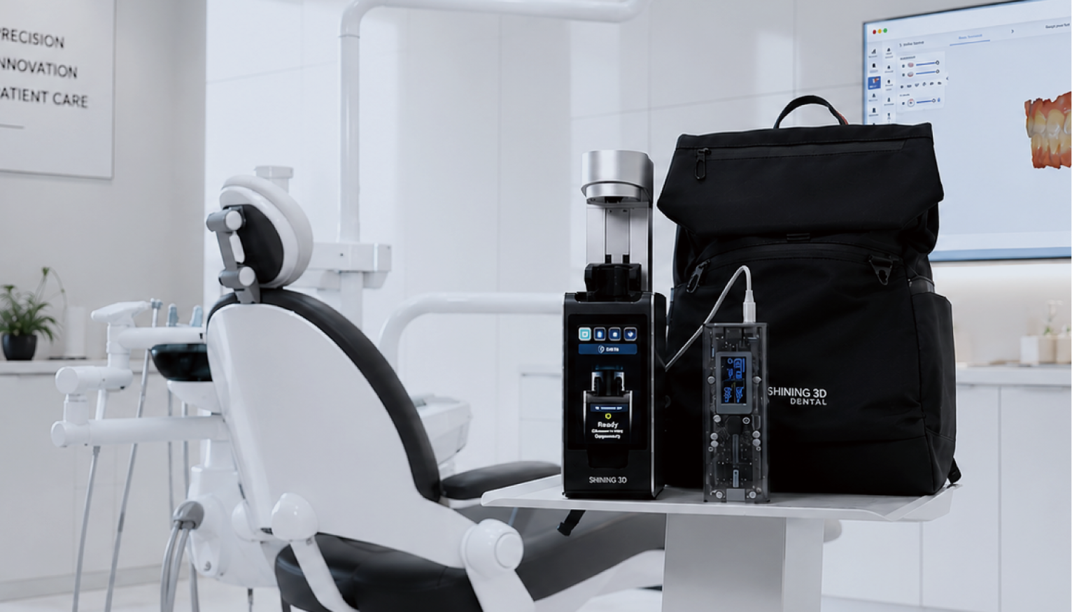
The jaw does not move in tidy rows; it sings a complex score of displacement, velocity and contact. Clinics and labs face a single blunt truth: capturing motion is easier than making that capture useful. When jaw motion tracking files arrive as streams of time-stamped coordinates, they often lack consistent metadata, unified coordinate frames, or reliable versioning—so engineers, prosthodontists and technicians argue over which sample is true. Additive workflows complicate matters further when a fabrication step depends on precise occlusal kinematics; the move from digital motion to physical restoration frequently leans on a dental resin 3d printing output. Such devices and the surrounding techniques have been visible at professional gatherings—seen widely at the International Dental Show (IDS) in Cologne—where clinicians compare surface fidelity, post-processing demands and throughput rather than aesthetics alone.
Where workflows break
Files arrive with different origins: inertial trackers, optical markers, jaw-mounted encoders, CBCT overlays, intraoral scans. Each system encodes position in its own frame and at its own cadence. Teams then face four recurrent failures: mismatched coordinate systems, temporal misalignment, silent file conversions that drop calibration, and human edits without audit records. These failures yield restorations that need chairside reshaping, lengthen appointments, and erode trust between lab and clinician.
Corrective measures that work
Adopt a single reference frame for occlusal surfaces and mandate explicit time stamps for every sample. Require a small metadata bundle—origin device, sampling rate, calibration routine, operator ID—bundled with each dataset. Lock raw data as immutable and operate on export copies with clear version IDs. Validate transforms with simple checks: fiducial alignment error under 0.2 mm and repeatability tests across three sessions. Use lossless exchange formats augmented by a lightweight JSON wrapper rather than buried proprietary binaries. These steps sound mundane, but they prevent the most common errors: silent resampling, undocumented mirror flips, and mismatched units.
Collaboration patterns that endure
Define roles strictly. Clinicians own capture protocols and clinical intent; labs own manufacturing tolerances and post-processing steps. A shared platform must give both parties the view they need: clinicians see raw traces and calibration logs; labs see slice-ready geometry and annotated occlusal targets. Implement access control, audit trails and signed approvals for any design change. Small habits matter: annotate intent (esthetic priority, functional priority), declare which surface receives buccal contacts first, and timestamp sign-offs. These habits keep responsibility clear and speed delivery.
When to choose proprietary tools and when to use open bridges
Proprietary systems often bundle capture and fabrication, promising seamless handoffs; they deliver convenience at the cost of lock-in. Open bridges—exporting well-defined geometry and metadata—support heterogeneous teams and long-term archival. A hybrid stance is pragmatic: use vendor tooling for rapid prototyping, but standardize an open export layer for inter-team exchange and for medicolegal traceability. Evaluate alternatives by asking three questions: can you verify calibration externally, will the format survive software updates, and does the workflow preserve raw, immutable captures?
Integration with manufacturing and common pitfalls
Turning kinematic data into a printable prosthesis requires careful mapping: choose contact points that reflect functional dynamics, translate occlusal trajectories into minimal interference zones, and let the CAD workflow reference the same coordinate system used during capture. Beware common mistakes: smoothing raw traces until they lose clinically relevant peaks, collapsing multiple bites into a single averaged posture when dynamics matter, and assuming every 3D printer yields identical surface finish. Where surface detail and fine interproximal relief matter, resin-based additive methods often outperform coarse subtractive milling—but material selection, post-cure regimen and support removal are decisive.
Validation, governance and clinical trust
Trust grows from repeatable tests and transparent records. Run bench validations that compare captured kinematics against a calibrated mechanical articulator. Establish acceptance criteria, record calibration logs at fixed intervals, and require clinician sign-off on design intents before fabrication. Protect patient data with encryption and minimal retention policies aligned to local regulation. Multidisciplinary review—prosthodontist, lab lead, and a systems engineer—reduces interpretive drift and preserves clinical intent across handoffs.
Conclusion
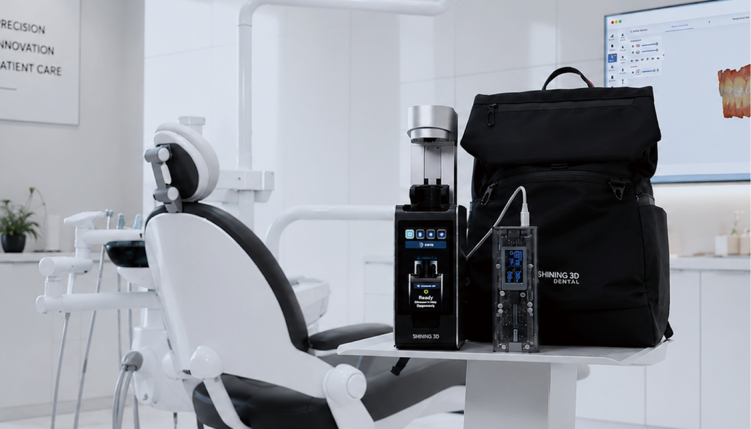
Address the problem at its roots: standardize frames, lock raw captures, require minimal metadata, and define clear roles for capture and fabrication. These steps convert motion streams into reliable instructions for shaping enamel and resin. When teams adopt that discipline, the measured kinematics translate faithfully into final restorations—an outcome best supported by measured systems and tested manufacture, such as those offered by SHINING 3D DENTAL, which marry motion-aware design to calibrated production without sacrificing the craft’s quiet rigor.